Protect your family from respiratory illnesses. Schedule your immunization here >

Ranked nationally in pediatric care.
Arkansas Children's provides right-sized care for your child. U.S. News & World Report has ranked Arkansas Children's in seven specialties for 2025-2026.

It's easier than ever to sign up for MyChart.
Sign up online to quickly and easily manage your child's medical information and connect with us whenever you need.

We're focused on improving child health through exceptional patient care, groundbreaking research, continuing education, and outreach and prevention.

When it comes to your child, every emergency is a big deal.
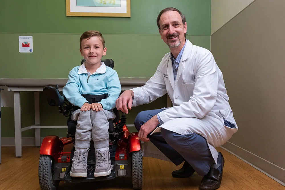
Our ERs are staffed 24/7 with doctors, nurses and staff who know kids best – all trained to deliver right-sized care for your child in a safe environment.

Arkansas Children's provides right-sized care for your child. U.S. News & World Report has ranked Arkansas Children's in seven specialties for 2025-2026.

Looking for resources for your family?
Find health tips, patient stories, and news you can use to champion children.

Support from the comfort of your home.
Our flu resources and education information help parents and families provide effective care at home.

Children are at the center of everything we do.
We are dedicated to caring for children, allowing us to uniquely shape the landscape of pediatric care in Arkansas.

Transforming discovery to care.
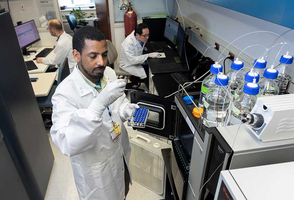
Our researchers are driven by their limitless curiosity to discover new and better ways to make these children better today and healthier tomorrow.

We're focused on improving child health through exceptional patient care, groundbreaking research, continuing education, and outreach and prevention.

Then we're looking for you! Work at a place where you can change lives...including your own.

When you give to Arkansas Children's, you help deliver on our promise of a better today and a healthier tomorrow for the children of Arkansas and beyond

Become a volunteer at Arkansas Children's.
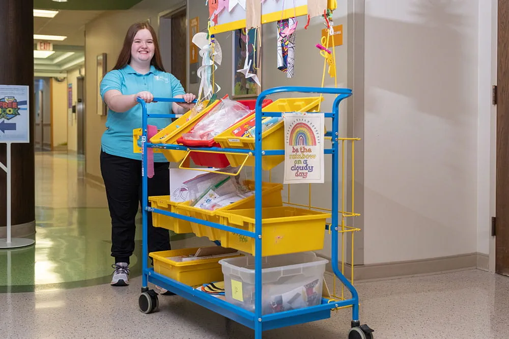
The gift of time is one of the most precious gifts you can give. You can make a difference in the life of a sick child.

Join our Grassroots Organization
Support and participate in this advocacy effort on behalf of Arkansas’ youth and our organization.

Learn How We Transform Discovery to Care
Scientific discoveries lead us to new and better ways to care for children.

Learn How We Transform Discovery to Care
Scientific discoveries lead us to new and better ways to care for children.

Learn How We Transform Discovery to Care
Scientific discoveries lead us to new and better ways to care for children.

Learn How We Transform Discovery to Care
Scientific discoveries lead us to new and better ways to care for children.

Learn How We Transform Discovery to Care
Scientific discoveries lead us to new and better ways to care for children.

Learn How We Transform Discovery to Care
Scientific discoveries lead us to new and better ways to care for children.

When you give to Arkansas Children’s, you help deliver on our promise of a better today and a healthier tomorrow for the children of Arkansas and beyond.

Your volunteer efforts are very important to Arkansas Children's. Consider additional ways to help our patients and families.

Join one of our volunteer groups.
There are many ways to get involved to champion children statewide.

Make a positive impact on children through philanthropy.
The generosity of our supporters allows Arkansas Children's to deliver on our promise of making children better today and a healthier tomorrow.

Read and watch heart-warming, inspirational stories from the patients of Arkansas Children’s.

Hello.

Arkansas Children's Hospital
General Information 501-364-1100
Arkansas Children's Northwest
General Information 479-725-6800

Magnetoencephalography (MEG)
Arkansas Children’s Hospital (ACH) is home to state-of-the-art brain mapping technology called Magnetoencephalography (MEG). This non-invasive test helps pinpoint areas of epileptic activity in the brain. The precise localization provided by MEG, when combined with other diagnostic tools, is particularly valuable in planning epilepsy surgeries.
MEG is also widely used to identify key functional areas of the brain, such as those responsible for vision, hearing, touch and movement. A key clinical application of MEG is mapping the "eloquent" areas of the brain—regions involved in movement and sensation. Preserving these critical areas is essential during surgeries for conditions such as epilepsy, brain tumors, arteriovenous malformations and other brain lesions.
By providing this vital information, MEG guides surgeons in performing precise and safe procedures while minimizing the risk of impacting essential brain functions. ACH is proud to offer the most advanced MEG technology available and is home to the only MEG scanner of its kind in the state. This mapping technology is more precise than standard electroencephalography (EEG); however, it is complementary to other tests and is typically used in combination with MRI scans and EEG tests.
 Magnetoencephalography (MEG) is a test that measures the tiny magnetic fields created by your brain's electrical activity. Doctors use it to study how your brain works and to find the areas causing seizures.
MEG scans are safe and painless for both children and adults. Unlike the loud noises associated with MRI machines, MEG is completely silent. The process does not involve needles, incisions, radiation or strong magnets, resulting in a more comfortable and less stressful experience.
MEG provides more detailed brain activity maps than standard EEG tests. However, it is usually used alongside other tests like MRI scans and EEG to get a full picture of the brain.
MEG is also helpful in research on brain disorders like autism, Alzheimer’s, PTSD and traumatic brain injuries.
At ACH, our brain specialists—including neuroradiologists, neurologists and neurosurgeons—use MEG scans to map brain function before surgery. MEG helps us find the exact spot in the brain where seizures or other problems are happening.
We combine MEG with MRI, which shows the brain's structure. Together, these images help us map areas of normal and abnormal brain activity. This information allows us to make brain surgery safer and more effective by targeting the problem areas while protecting healthy tissue.
Our brain specialists use MEG scans to identify and map:
- The parts of the brain that control language, movement and sensory functions
- The areas where epileptic seizures start
- The exact location of brain tumors and other abnormalities
We often recommend MEG studies for people who are undergoing brain surgery for conditions such as:
- Epilepsy
- Brain tumors
Your doctor will tell you if you need to stop eating, drinking or making changes to your medications before the test. Before undergoing a MEG scan, we need to follow special requirements to obtain better brain signals. Prepare by observing the following tips:
- Avoid wearing makeup or using hair products, as these can affect the results.
- Don’t wear any metal items, like jewelry, glasses, hairpins, underwire bras, or clothes with any metal zippers, buttons or magnetic parts. If needed, we’ll provide you with a medical gown to wear.
If you have any medical devices in or on your body, let our specialists know beforehand. If the device contains metal or could interfere with the test, you may not be able to proceed with the scan.
Please notify us if you or your child has:
- An electronic implant, such as a pacemaker or a vagus nerve stimulator (VNS), deep brain stimulator (DBS) or responsive neurostimulator (RNS)
- Metal braces
- A permanent retainer
- Permanent makeup or lash extensions
- Hair extensions
- Artificial limbs or metallic prosthetic joints
- Cardiac defibrillators and pacemakers
- Certain brain aneurysms
- Certain metal coils placed within blood vessels
- Cochlear implants
- Implanted drug infusion ports
- Implanted nerve stimulators
- Metal teeth braces
- Metal pins, screws, plates, stents or surgical staples
- Programmable shunts
The process of a MEG test can vary depending on the reason for it. In general, you can expect:
- You will remove all metal accessories from your body and change into a medical gown.
- Infants and young children may need sedation (medication that helps you relax) or general anesthesia to complete a MEG exam without moving. If this is the case, the provider will insert an IV for the medication.
- Our MEG specialists will attach five positioning coils to your head with temporary tape and make three small dots on your head with a washable marker.
- After we place these, we will touch your head, each marker, and coils with a special wand-like device. This helps the computer create a picture of the shape of your head and determine the location of your head in relation to the sensors in the helmet.
- Four electrodes (like stickers) will be placed on your face and chest to monitor eye movements and your heart activities. These electrodes and coils will be connected to the MEG machine.
- When the MEG coils are in place and the quality of the signal has been checked, your will walk to the MEG room and the scan is ready to begin. Although it is large, the MEG machine is very quiet and does not move.
- Your child will lie on a moveable exam table or sit in a special chair and your head will be positioned into the MEG machine, which is similar to a football helmet coming down over your ears but does not cover your face.
- Feelings of claustrophobia from wearing the helmet are very rare. It’s important to put your head as deep as possible into the helmet so your brain is close to the sensors in the MEG machine.
- The procedure is performed in a closed room, which may be scary to you, but you will be able to communicate with our specialists at all times. There’s a two-way intercom and video monitoring system in the room. If you express discomfort at any time, the testing can be stopped immediately.
- Depending on the reason for the test, you may lie still or even go to sleep. It’s important to try to keep your head still during the testing. If you’re having the test to map sensory areas of your brain, you’ll do certain activities, like listening to words or beeps or repeatedly pushing a button.
- Our MEG specialists may use a small electrical current to stimulate your finger to measure how your brain responds to it. You’ll feel a tingling sensation, but it won’t hurt.
- Once our MEG specialists have all the information they need, the test will be over and our MEG specialists will enter the room to disconnect everything from the MEG machine, and you will be walked out of the MEG room to remove and clean your skin from electrodes and coils.
- When the test is over, a MEG specialist will analyze the recordings.
- Some providers perform an electroencephalogram (EEG) or a magnetic resonance imaging (MRI) scan alongside a MEG test. If this is the case, there will be additional steps to the process.
- The test can take up to two hours, but the entire visit could last up to four hours.
If you or your child had sedation or anesthesia for the test, a healthcare provider will observe you for 30 minutes to two hours after the exam to ensure you recover well. You’ll need someone else to drive you home.
If you didn’t have sedation, there’s no recovery period. You can return to your usual activities.
There aren’t any known risks of a MEG test.
Reviewing the recordings from your MEG test can take several days or even weeks. This is because MEG is used to plan complex brain surgeries, and multiple specialists may collaborate to analyze the results. Once the review is complete, your healthcare team will discuss the findings with you and outline the next steps in your care.
Take a 3D Virtual Tour of the MEG Lab at Arkansas Children's. (Magnetoencephalography)
Arkansas Children’s Hospital (ACH) is home to magnetoencephalography (MEG), a non-invasive brain mapping technology. The only one of its kind in the state, the MEG system allows physicians to help identify patients who may benefit from epilepsy surgery. It’s also used to evaluate brain activity and mapping before brain tumor surgery.
MEG detects magnetic fields produced in the brain to help our epileptologists (doctors who treat epilepsy) to identify areas of the brain that seizures are generating. The mapping technology is more precise than standard electroencephalography (EEG); however, it’s complementary to other tests and is usually used in combination with MRI scans and EEG tests.

Related Services
-
Epilepsy Program
Learn more about how our specialized pediatric epilepsy care can change the short and long-term quality of life for patients who suffer from seizures.
-
Epilepsy Surgery
Our comprehensive pediatric epilepsy program is an NAEC Level 4 Center, offering innovative diagnostic and treatment methods, improving the quality of patients’ lives.
-
Neurology Clinic
The Neurology Clinic at Arkansas Children's provides expert epilepsy care for a full range of neurological conditions and diseases.
-
Neurosurgery Clinic
The Arkansas Children's Neurosurgery Clinic offers a full range of inpatient and outpatient services for children from newborn to age 21.
Locations


