


Hello.

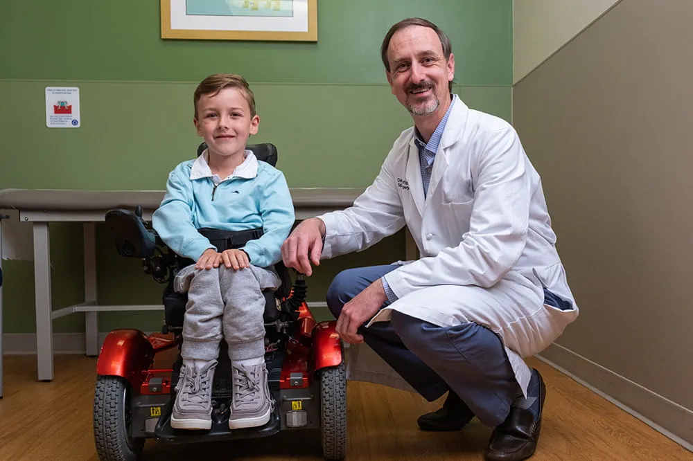
Arkansas Children's Hospital
General Information 501-364-1100
Arkansas Children's Northwest
General Information 479-725-6800

Fetal Echocardiogram
What is a fetal echocardiogram?
A fetal echocardiogram, also called a fetal echo, is a type of ultrasound that looks at your baby’s heart before they are born. A fetal echo uses sound waves to show the structure of your baby’s heart and how it is working from many views and perspectives. It can show details such as heart rate, heart rhythm, chamber sizes and cardiac valves. A fetal echo is an important tool to help find congenital heart problems before your baby is born.
Why would I need a fetal echo?
- You have a family history that puts you at high risk of having a baby with congenital heart defect (CHD)
- You have had another child with a CHD
- You have had an abnormal result from another test
- You are taking a medicine that may cause a congenital heart defect
- You have certain health conditions such as diabetes or an autoimmune disorder
When is a fetal echo done?
The screening is recommended to take place by 24 weeks of pregnancy.
What happens during a fetal echo?
A fetal echocardiogram is a painless test. It is much like other ultrasounds you may have had during your pregnancy. For the test, you will lie down on the exam table and have gel put on your stomach. This allows the sound waves to travel better from your uterus. The person doing the echo will use a special wand, called a transducer, to get images of your baby’s heart. They will move the transducer across your stomach to get images from different angles. The test usually takes about an hour, but it can vary depending on your baby’s position.